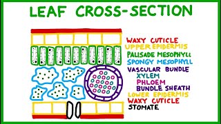
This makes it hydrophobic, meaning that it repels water. Its hydrophobic nature is what causes water droplets to bead on a leaf rather than soaking in. Just underneath the waxy cuticle is the second layer, which is called the upper epidermis. The cells of the upper epidermis are very closely packed together.
The upper epidermis has mostly protective functions; it allows light to come in, but also prevents water loss in conjunction with the waxy cuticle. The third layer moving down from the top is called the palisade mesophyll. If we were to look very closely at the cells of the palisade mesophyll, we would notice that each of them has a nucleus and also has a very large vacuole for water storage. However, arguably the most important thing about the palisade mesophyll is the fact that these cells are full of chloroplasts.
This makes the third layer the palisade mesophyll the main site of photosynthesis in a leaf. Photons of light will shine down through the waxy cuticle and the upper epidermis, which are both mostly clear, and allow photosynthesis to take place in the chloroplast of the palisade mesophyll. The fourth layer from the top underneath the palisade mesophyll is called the spongy mesophyll. Now, before you start drawing, please leave this space in the box blank for a minute.
The cells of the spongy mesophyll are a little bit more irregular in shape, but if we look a little bit more closely, we can see that they actually do contain some chloroplasts. This means that they can do some photosynthesis; not as much as the palisade mesophyll. The cells of the spongy mesophyll are also spaced pretty far apart, and the area in between them is called the intercellular space. In this space is usually water and oxygen.
Now we're going to fill up this empty space right here on the right and we're going to fill it with a structure called the vascular bundle. If we go back to our original leaf cross-section, the vascular bundle is going to be this area right here, what we would originally called the stem of the leaf, but we're not going to call it a stem. The vascular bundle is round and is responsible for moving products around the leaf. Just like you have a water pipe that brings water into your house and a sewage pipe that removes the wastewater, the vascular bundle works similarly.
The outside coating of the vascular bundle is made up of a large group of closely packed cells; these cells are called bundle sheath cells and they keep all of the vasculature together. Inside the bundle sheath cells is a tightly packed group of tubules called xylem and phloem. The purpose of the xylem is to bring the water up from the roots of the plant in order to nourish the leaves. Remember that water is a key component in photosynthesis.
After photosynthesis, the phloem will bring the glucose molecules all around the plant to all the parts that need it. Even though these two types of tubules are doing different things, they are right next to each other which makes for a very efficient system. Don't forget that even though there are multiple types of structures within the vascular bundles, the vascular bundle only refers to the structure as a whole. There are still three types of tissues; phloem and xylem and bundle sheath cells.
Now we're going to move on to drawing the lower layers of the leaf and some of these will look pretty familiar. Just underneath the vascular bundle and the spongy mesophyll is another layer of epidermis. However this is called the lower epidermis because it's on the lowest part of the leaf. Just like the upper epidermis, the lower epidermis has cells that are packed very closely together in order to prevent water loss.
Finally, underneath the lower epidermis we have one more waxy cuticle, just like on the top. It helps to prevent water loss. So now we have a nice picture that shows all of the different layers of the leaf, but the problem is that this current leaf as it stands is completely watertight, it doesn't allow for any gas exchange. Remember that in photosynthesis photons of light shine onto leaves, we add carbon dioxide and water, and then after the light and dark reactions, we produce 2 PGAL which become glucose and also oxygen.
But, our leaf as it currently stands can't undergo any gas exchange, so we're going to need to introduce a couple of modifications to the bottom layer of our leaf. We've now zoomed in on the lower waxy cuticle and the lower epidermis of the leaf, and we can also see a little bit of the vascular bundle up top. At the very bottom of the leaf, we're going to add a small opening to permit gas exchange. This structure here is called a stomate, and if we were to zoom out a little bit we would notice at the bottom layer the leaf actually has a fairly sizable number of them.
The plural of stomate is stomata. This opening allows for carbon dioxide gas to flow in, oxygen gas to flow out, and water vapor to also flow out. However, if we left it open all the time, the leaf would rapidly dry out. These two structures making up either side of the stove mate are called guard cells; the guard cells can pinch together in order to open or close the stomate as necessary.
Here you can see what the leaf actually looks like if you took a cross-section and examined it under the microscope after giving it a little dye. The waxy cuticle is very hard to see because it's so thin, but if you look at the area circled in red you can see a little hint of it. This particular picture shows this vascular bundle is being called the vein, which is also correct, but for our intents and purposes, we will call it a vascular bundle. Finally here is an unlabeled cross-section of a leaf, again under a microscope.
See if you can identify all of the structures labeled here, but please ignore this one. Here's the answer key for this particular cross section; even though all the layers were originally the same color it's pretty easy to tell them apart by cell shape. The waxy cuticle is challenging to see, but the epidermis cells tend to be square shaped and packed very closely together. In the palisade mesophyll, which is outlined in green, the cells are what we call columnar, they're very long and skinny.
In the spongy mesophyll, the cells are easy to pick apart because they have a large space in between them and they're also more irregularly shaped. The lower epidermis and the waxy cuticle on the bottom look very similar to what's on the top. Stovmata are easy to see because they look a little bit different than the lower epidermis cells; you only find stomata on the lower surface of the leaf and never on the top. Finally, the vascular bundle is distinct because the cells are very closely packed together and they tend to be more rounded shape.
For our intents and purposes you don't have to be able to tell apart xylem and phloem; just know that they're both there and what the functions of both of them are. That's it! See you all in class!.
No comments:
Post a Comment